
Harnessing the Potential of AI in Oncology: Current Applications and Future Outlook
Introduction to AI
Artificial Intelligence (AI) is the field of computer science that strives to present machines with the ability to simulate/emulate human like intelligence and cognition. AI systems are designed to learn from data and output its decision. The intelligence comes from the set of instructions called algorithm developed by humans to recognize patterns, make decisions, and perform tasks that typically require human intelligence, such as understanding natural language, solving complex problems, and making recommendations. This transformative technology has far-reaching applications, from self-driving cars and virtual assistants to financial forecasting and precision oncology in medical diagnostics and treatment of cancer.
Artificial Intelligence (AI), Machine Learning (ML) and Deep Learning (DL) are closely related as shown in the figure below. AI is the broader spectrum that focuses on creating intelligent machines whereas ML is a subset of AI that focuses on developing algorithms and models that can learn from data. Instead of explicitly programming rules, ML systems use data to learn patterns and make predictions or decisions. ML encompasses a wide range of techniques, including regression, classification, clustering, and reinforcement learning. DL is a subfield of ML that emphasizes the use of neural networks, specifically deep neural networks with multiple layers (hence "deep"). These deep neural networks are particularly suited for tasks such as classification and regression involving unstructured data, such as images, audio, and text. DL has achieved remarkable success in various AI applications, including computer vision, speech recognition, and natural language processing.

Fig 1: Artificial intelligence, machine learning and deep learning
Source: Nadia BERCHANE (M2 IESCI, 2018)
Why is AI gaining popularity now?
AI's popularity has surged in recent years, primarily due to significant advancements in both data availability and computing performance. In recent times, the digital age has led to an unprecedented era of data generation. Vast amounts of structured and unstructured data are continuously being produced by various sources, including social media, sensors, e-commerce transactions, hospital data and more. Many institutions and organizations have made their data available to the public, which has further expanded the resources accessible for AI research and development. This wealth of data serves as the fuel for training and improving AI models. On the other hand, advances in hardware, particularly the development of powerful GPUs (Graphics Processing Units) and TPUs (Tensor Processing Units), have made it possible to train and run complex AI models much more efficiently. AI algorithms, especially DL models, can take advantage of parallel processing on these advanced hardware platforms, dramatically reducing training times with higher accuracy sometimes surpassing human intelligence.
Application of AI in different fields
Artificial Intelligence (AI) has found applications in various fields, transforming industries, and improving efficiency, decision-making, and automation. Specifically in healthcare for Diagnosis and Disease Prediction; objectives like medical image analysis (like X-rays and MRIs, Digitized pathology slides) and patient data analysis to assist doctors in diagnosing diseases such as cancer, diabetes, and heart conditions is handled with higher accuracy. In addition, it can be used in accelerating drug discovery and provide robotic services and chatbot support for patient care. Apart from health care, AI has wider applications in other domains to name a few like, retail, finance, education etc.
AI in oncology: Application of AI in diagnosis, prognosis, and treatment plan
Case-study (Diagnosis Task): AI based automated nuclei segmentation for differentiating benign and malignant cancer.
The introduction of advanced digital slide scanners for whole slide images has renewed interest in pathology image analysis. Researchers [1] have applied an automated nuclei segmentation method developed specifically for hematoxylin and eosin (H&E) stained breast cancer histopathology images from digital slides. This method consists of four main steps: color unmixing and morphological operators for pre-processing, marker-controlled watershed segmentation at multiple scales with different markers, post-processing to eliminate false regions, and merging results from multiple scales. The method was tested on 21 breast cancer cases and evaluated for detection accuracy (sensitivity and positive predictive value) and segmentation accuracy (Dice coefficient). Results show high sensitivity and positive predictive values with most segmentations having a Dice coefficient larger than 0.8.

Fig 2: AI based automated nuclei segmentation for differentiating benign and malignant cancer.
Case-study (Prognosis task): Survival analysis on whole slide images using deep learning.

Fig 3: deep learning model architecture
In this study researchers [2] aimed to address the variability in breast cancer grading by training a deep learning model to assess tumor grade using whole-slide histopathology images. The model was trained on data from 706 young invasive breast cancer patients and evaluated using an independent test set of 686 patients. It achieved an 80% accuracy and a Cohen's Kappa of 0.59, outperforming expert pathologists in distinguishing low/intermediate from high-grade tumors. Survival analysis indicated significantly different overall and disease/recurrence-free survival rates between the model-predicted groups. Although this trend weakened after adjusting the clinicopathologic features and molecular subtypes, significantly the model distinguishes between low/intermediate and high-grade tumors and finds a trend in the survival of the two predicted groups.
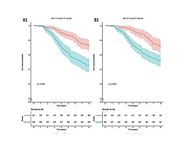
Fig 4: Survival analysis graphs.
Case-study (Treatment task): Estimate the treatment response of neo-Adjuvant treated breast cancer patients based on identifying cellularity from H&E images.
This paper [3] presents a method to automatically assess Tumor Cellularity (TC) in Neo-Adjuvant treatment (NAT) breast cancer patients using morphological features extracted from histological images. TC is crucial for measuring therapy efficacy. The approach combines traditional computer vision techniques with Machine Learning (ML) algorithms, leveraging key morphological parameters. The method's accuracy, validated against pathologist scores, improved compared to other ML methods and approached the performance of deep learning, suggesting its potential utility in cases with limited training data where deep learning may not be feasible.

Fig 5: DL architecture for estimating the responder vs non-responders.
What is the future of AI in oncology?
There has been a constant effort from the genomic side to precision oncology that relies on genetic and molecular profiling of a patient's tumor to identify specific mutations and characteristics. This information is used in the selection of targeted therapies and treatment strategies, maximizing effectiveness, and minimizing side effects. One prominent example would be to identify epidermal growth factor receptor (EGFR) mutation in non-small cell lung cancer (NSCLC). Identifying EGFR mutations in NSCLC is highly significant and has profound implications for patient care and treatment outcomes such as improved survival rate, reduced side effect, monitoring resistance to therapies. It is well known that NGS based methods are looking at molecular profiles, imaging methods both invasive and non-invasive techniques, would pick up morphological features differentiating w.r.t the size and shape of the cells. For example, in the case of EGFR, elongated or spindle-shaped cancer cells might contribute significantly to identifying the mutation. In addition, cancerous nuclear enlargement or pleomorphism would also be a prominent feature in detection the mutation.
It is evident that one modality, either NGS or Imaging techniques will not comprehend totally in understanding the dynamics of cancer biology. Towards this end there is a pressing demand to integrate different modalities of data to harness the power of AI in treating cancer more effectively. Ultimately, the goal is to improve treatment outcomes and enhance the quality of care for cancer patients.
The future of AI in oncology holds on combination of multiple “omics” by integrating data from imaging, genomics, proteomics and clinicopathological. We believe that a multi-omics-based AI system as shown in Fig 6 would help to collate the knowledge from all systems in silos. Such an AI system will be capable of understanding the comprehensive complex biological system of the cancer and in turn will suggest and guide clear, precise, and better treatment for the cancer patients. This interdisciplinary approach is highly important in advancing our knowledge of cancer biology and improving healthcare with optimal treatment for cancer patients.

References:
Veta M, van Diest PJ, Kornegoor R, Huisman A, Viergever MA, Pluim JPW (2013) Automatic Nuclei Segmentation in H&E Stained Breast Cancer Histopathology Images. PLoS ONE 8(7): e70221.
Wetstein, S.C., de Jong, V.M.T., Stathonikos, N. et al. Deep learning-based breast cancer grading and survival analysis on whole-slide histopathology images. Sci Rep 12, 15102 (2022).
Ortega-Ruiz, M. A., Karabağ, C., Garduño, V. G., & Reyes-Aldasoro, C. C. (2020). Morphological estimation of Cellularity on Neo-adjuvant treated breast cancer histological images. Journal of Imaging, 6(10), 101.
Fig 6, Genetics abnormalities of different cancer Andres F. Cardona on X: "GENETICS ABNORMALITIES OF DIFFERENT CANCERS https://t.co/qEXRQp097I" / X (twitter.com)
Authors:
Anand Ulle
Data Scientist Imaging
OneCell Diagnostics Private Limited
https://www.linkedin.com/in/anand-ulle-58777747/
P M Shivamurthy
Data Scientist Imaging
OneCell Diagnostics Private Limited
https://www.linkedin.com/in/pmshiva/